UEG's content platform for gastroenterology education bringing recordings, courses, guidelines, and more together in one place.
Trending on Gutflix
Rate for better recommendations!
Your feedback helps us show you more of what you like and less of what you dislike!

Log in to continue.
This content is part of Gutflix. Log in with your myUEG account, or create one free, to watch it.
Not sure what you can access? Learn more about account types.
RADIOLOGICAL REMISSION IN PERIANAL FISTULIZING CROHN’S DISEASE: BASELINE MRI PREDICTORS AND IMPACT ON CLINICAL OUTCOMES
Sarra Laabidi 1, Amal Ghamougui 1, DONIA GOUIAA 1, sahar chtioui 1, Nadia Ben Mustapha 1, Meriem Serghini Ep Ghachem 1, Asma Labidi 1, Monia Fekih 1
Affiliations
1 Rabta Hospital, Tunis, Tunisia
Log in to continue.
This content is part of Gutflix. Log in with your myUEG account, or create one free, to watch it.
Not sure what you can access? Learn more about account types.
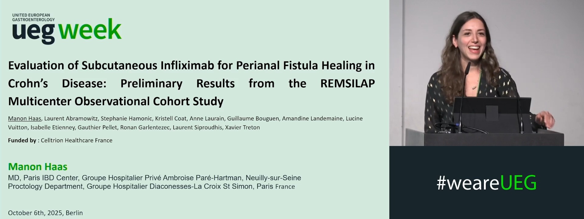
EVALUATION OF SUBCUTANEOUS INFLIXIMAB FOR PERIANAL FISTULA HEALING IN CROHN’S DISEASE: PRELIMINARY RESULTS FROM THE REMSILAP MULTICENTER OBSERVATIONAL COHORT STUDY
Manon Haas 1, Laurent Abramowitz 2, Stephanie Hamonic 3, Amandine Landemaine 3, Guillaume Bouguen 3, Anne Laurain 2, Lucine Vuitton 4, Isabelle Etienney 5, Gauthier Pellet 6, Kristell Coat 3, Ronan Garlantezec 3, Laurent Siproudhis 3, Xavier Treton 1
Affiliations
1 Institut des MICI - Clinique Ambroise Paré Hartmann, Neuilly sur Seine, France
2 CHU Bichat, Paris, France
3 CHU Pontchaillou, Rennes, France
4 Besançon university hospital gastroenterology, Courbevoie, France
5 Diaconesses-Croix St Simon Hospital, Paris, France
6 CHU Bordeaux, Bordeaux, France

Log in to continue.
This content is part of Gutflix. Log in with your myUEG account, or create one free, to watch it.
Not sure what you can access? Learn more about account types.
MULTI-OMICS ANALYSIS OF PERIANAL FISTULIZING CROHN'S DISEASE REVEAL NOVEL THERAPEUTIC TARGETS
Affiliations
1 Washington University in St. Louis, St. Louis, United States