UEG's educational platform for gastroenterology education bringing recordings, courses, guidelines, and more together in one place.
Most popular content
Rate for better recommendations!
Your feedback helps us show you more of what you like and less of what you dislike!
Log in to access this content.
Free for all myUEG account holders. Your access level is set automatically based on your occupation. Medical professionals get full access to all content. If you are a non-medical user, you can only access UEG Week content from congresses you attended.
Not sure what you can access? Learn more about account types.

EFFECT OF UPFRONT PRECUT TECHNIQUES (PAPILLOTOMY VERSUS FISTULOTOMY) WITHOUT ATTEMPTING STANDARD BILIARY CANNULATION ON POST ENDOSCOPIC RETROGRADE CHOLANGIOGRAPHY (ERC) PANCREATITIS (PEP) RATE AMONG HIGH-RISK COHORT FOR PEP UNDERGOING ERC- A RANDOMIZED CO
Harshal Mandavdhare 1, Arpit Sasani 1, ABHISHEK YADAV 1, Jimil Shah 1, VANEET JEARTH 1, Vishal Sharma 1, Harjeet Singh 1, Pankaj Gupta 1, Amol Patil 1, Saroj Kant Sinha 1
1 Post Graduate Institute Of Medical Education And Research, Chandigarh, Chandigarh, India
Conference
Topics
Submission format
Citation
Published
Log in to access this content.
Free for all myUEG account holders. Your access level is set automatically based on your occupation. Medical professionals get full access to all content. If you are a non-medical user, you can only access UEG Week content from congresses you attended.
Not sure what you can access? Learn more about account types.
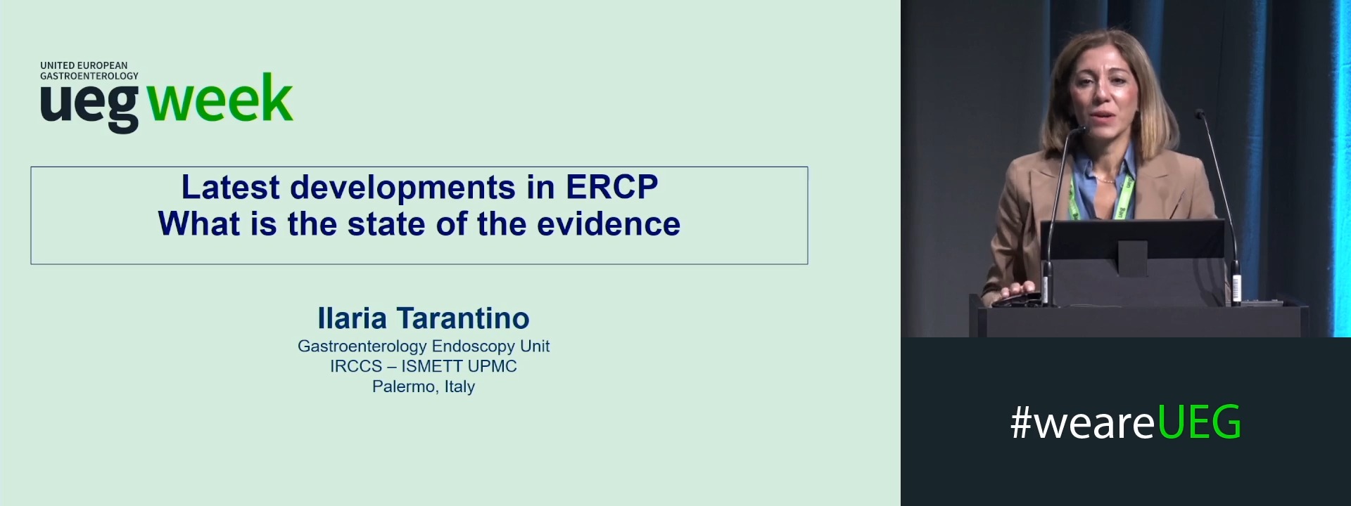
Latest developments in ERCP: What is the state of the evidence?
1 ISMETT, Palermo, Italy
Event
Citation
Published
Log in to access this content.
Free for all myUEG account holders. Your access level is set automatically based on your occupation. Medical professionals get full access to all content. If you are a non-medical user, you can only access UEG Week content from congresses you attended.
Not sure what you can access? Learn more about account types.
Panel discussion: Autoimmune disease of liver and bile ducts
Event
Topics
Citation
Published
Log in to access this content.
Free for all myUEG account holders. Your access level is set automatically based on your occupation. Medical professionals get full access to all content. If you are a non-medical user, you can only access UEG Week content from congresses you attended.
Not sure what you can access? Learn more about account types.
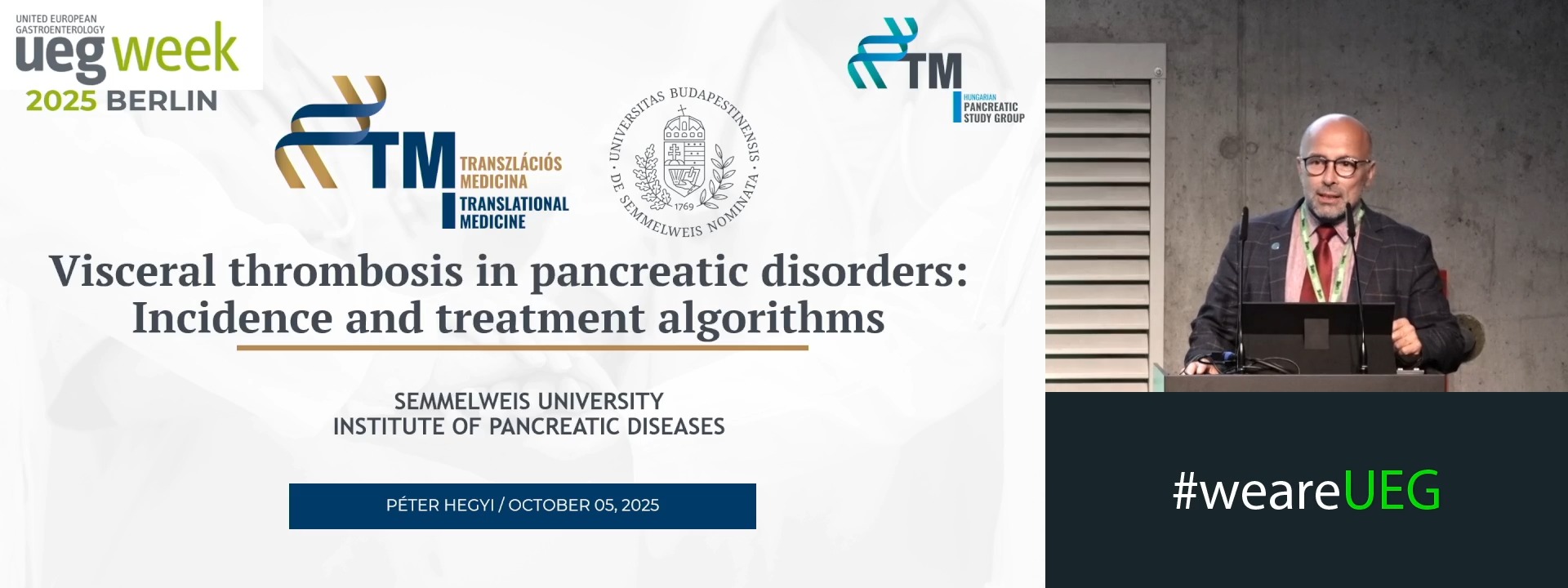
Visceral thrombosis in pancreatic disorders: Incidence and treatment algorithms
1 University of Pecs, Pecs, Hungary
Event
Topics
Citation
Published
Log in to access this content.
Free for all myUEG account holders. Your access level is set automatically based on your occupation. Medical professionals get full access to all content. If you are a non-medical user, you can only access UEG Week content from congresses you attended.
Not sure what you can access? Learn more about account types.
A RARE CASE OF PARANEOPLASTIC PEMPHIGUS AND BIOPSY-RELATED TUMOR SEEDING IN HEPATOCELLULAR CARCINOMA
Carolina Palma 1, Ivo Mendes 1, Francisco Vara-Luiz 1, Irina Mocanu 1, Gonçalo Nunes 1, Inês Costa Santos 1, Marta Patita 1, Unknown Unknown 1, Cristina Fonseca 1
1 Hospital Garcia de Orta, Almada, Portugal
Conference
Topics
Submission format
Session
Published
Log in to access this content.
Free for all myUEG account holders. Your access level is set automatically based on your occupation. Medical professionals get full access to all content. If you are a non-medical user, you can only access UEG Week content from congresses you attended.
Not sure what you can access? Learn more about account types.
WHEN ENDOSCOPIC SPHINCTEROTOMY IS NOT ENOUGH : EFFICACY AND SAFETY OF DILATION-ASSISTED STONE EXTRACTION FOR CHALLENGING BILIARY STONES: A TERTIARY CENTER EXPERIENCE
Oussama Nacir 1, Fatima Ezzahra Lairani 1, Adil Ait Errami 1, Sofia Oubaha 1, Zouhour Samlani 2, Khadija Krati 1
1 Mohammed VI University Hospital, Marrakech, Morocco|||Faculty of Medicine and Pharmacy, Cadi Ayyad University, Marrakech, Morocco
2 Mohammed VI University Hospital, Marrakech, Morocco
Conference
Topics
Submission format
Session
Citation
Published
Log in to access this content.
Free for all myUEG account holders. Your access level is set automatically based on your occupation. Medical professionals get full access to all content. If you are a non-medical user, you can only access UEG Week content from congresses you attended.
Not sure what you can access? Learn more about account types.
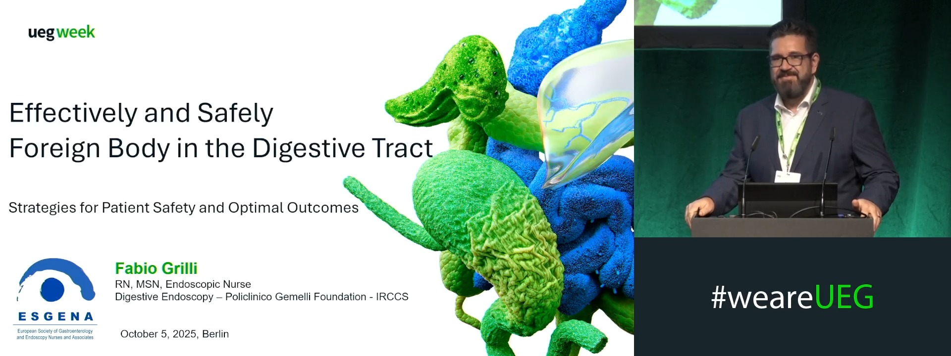
Effectively and safely- foreign body in the digestive tract
1 Fondazione Policlinico Universitario Agostino Gemelli IRCCS, Roma, Italy
Event
Topics